Overview
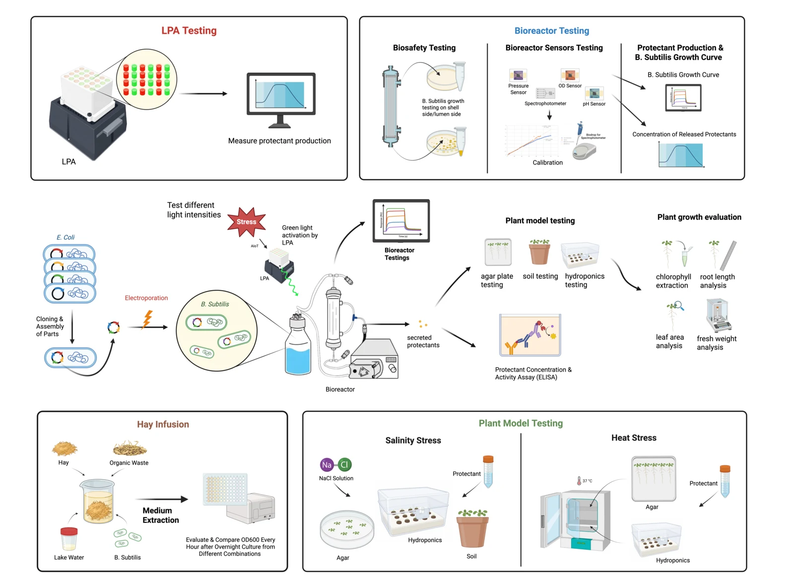
ReLEAF makes plant protectants in Bacillus subtilis and tests them on Arabidopsis under salt and heat stress. The wet lab work runs in four stages, and each hands its product to the next: cloning builds the constructs, protein production gets the protectant out of the culture and measures its activity, the plant assays test it, and the bioreactor sheets calibrate the instruments that watch the culture grow.
Protocols
Molecular cloning
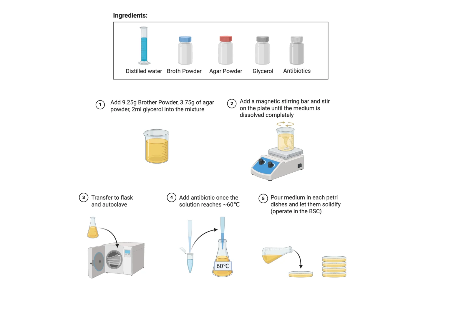
Culture agar preparationPrepare and make agar plates to cultivate bacteria.
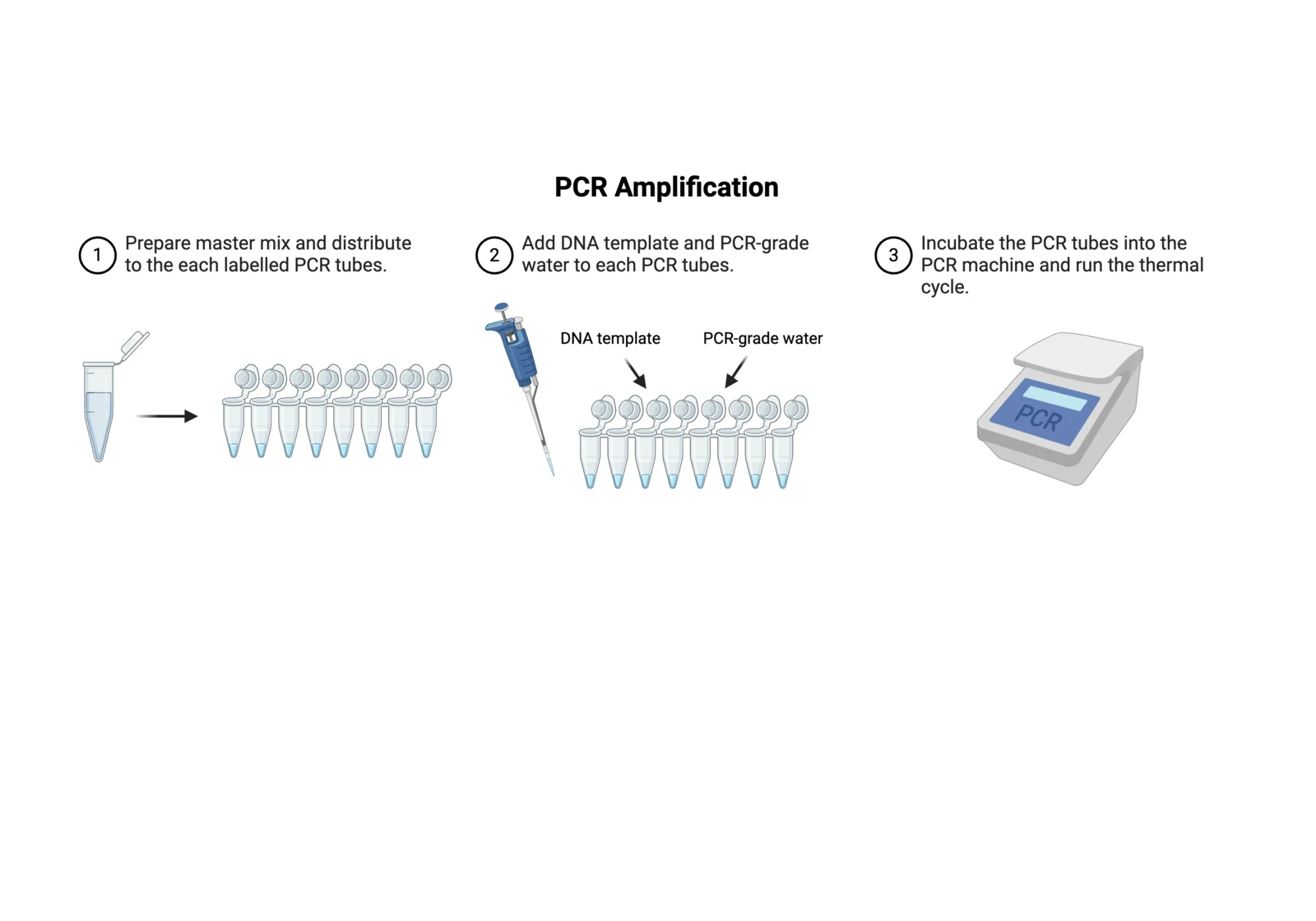
PCR amplificationA nucleic acid amplification technique to rapidly produce copies of DNA segments from a small initial sample.
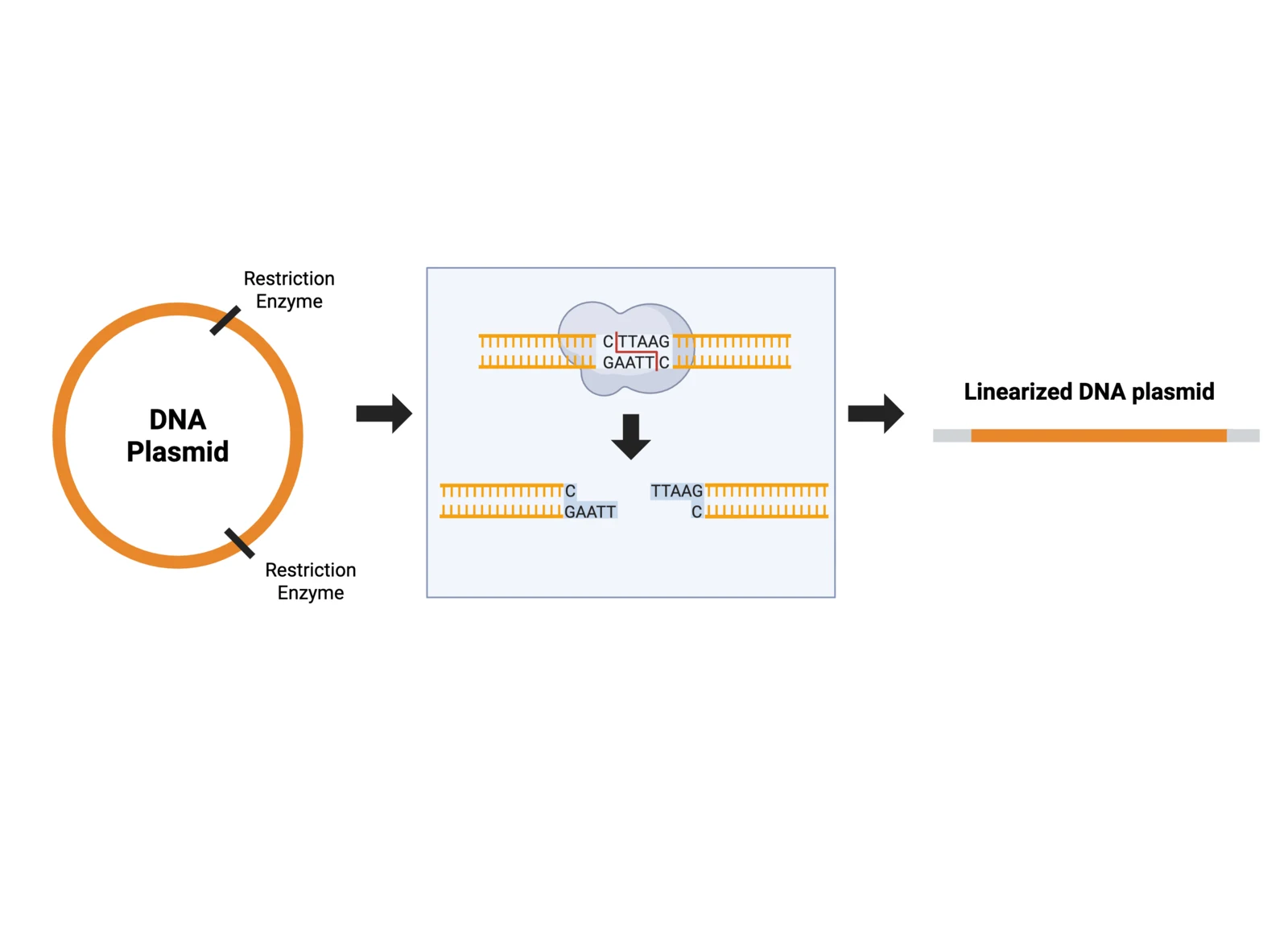
Restriction enzyme digestionLinearize purified plasmids with restriction enzymes to verify their sizes.
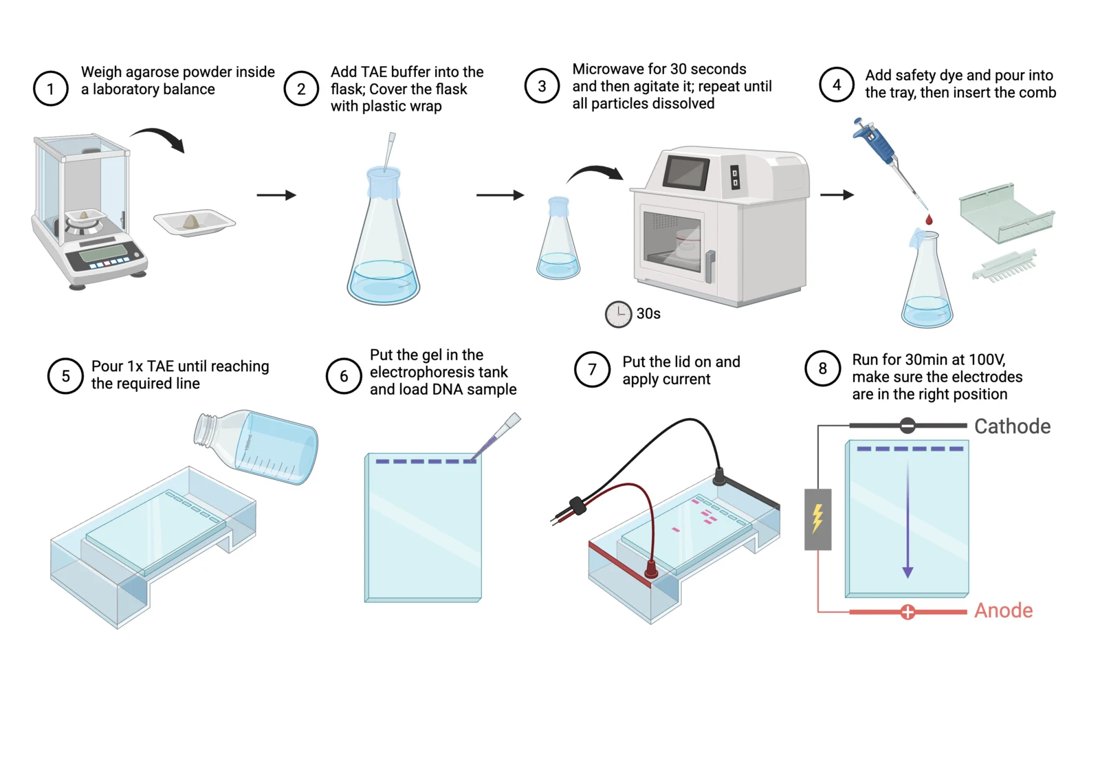
DNA gel electrophoresisResolve specific DNA fragments to allow visual verification and confirmation of product band sizes.
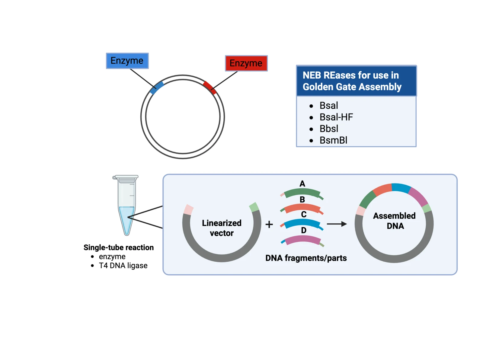
Golden Gate assemblyClone and assemble multiple core genetic parts of the light-controlled optogenetic circuit.
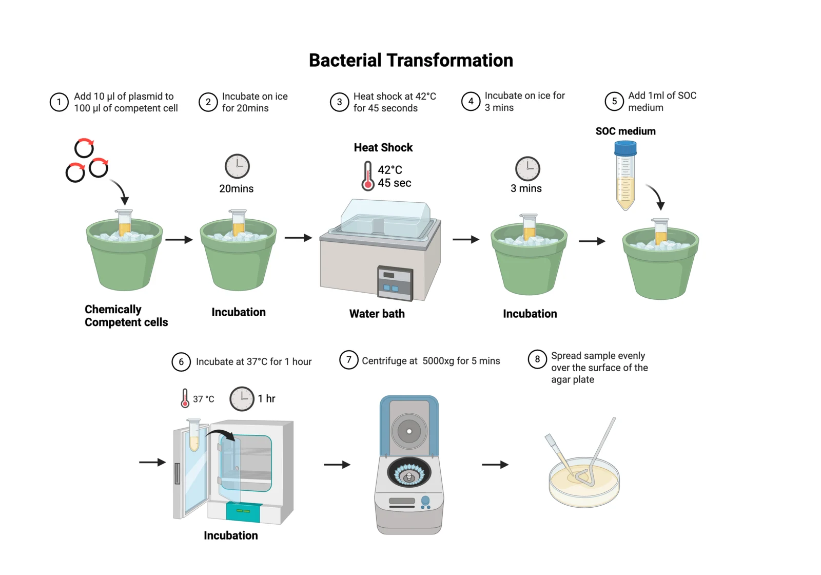
E. coli transformationIntroduce foreign plasmid DNA in competent cells in order to replicate plasmids in E. coli.
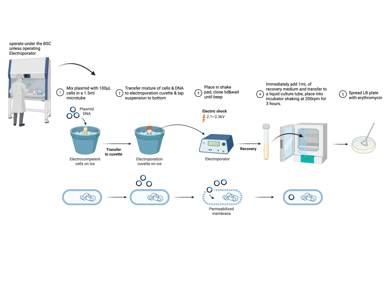
Electroporation into B. subtilisIntroduce foreign plasmid DNA in competent cells through exponential electrical flow in order to replicate plasmids in B. subtilis.
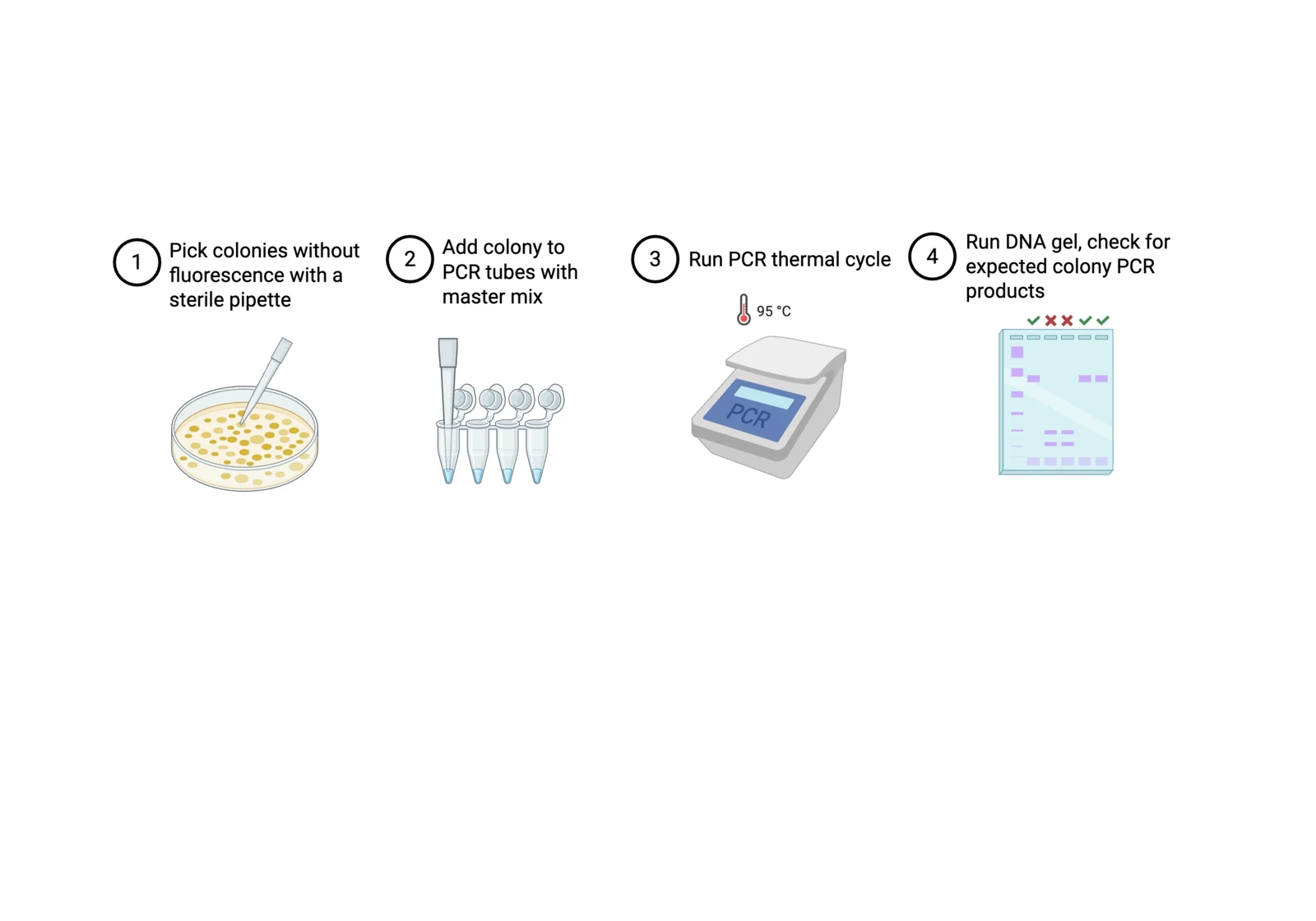
Colony PCRRapidly screen individual bacterial colonies to verify the presence of the desired DNA insert or recombinant construct using the colony as the PCR template.
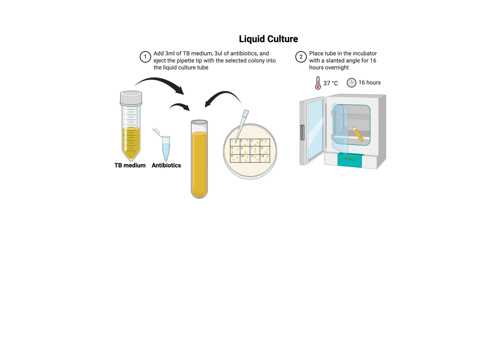
Liquid bacterial cultureGrow bacteria such as E. coli or B. subtilis in nutrient-rich broth, allowing rapid multiplication in a suspended, aerated environment.
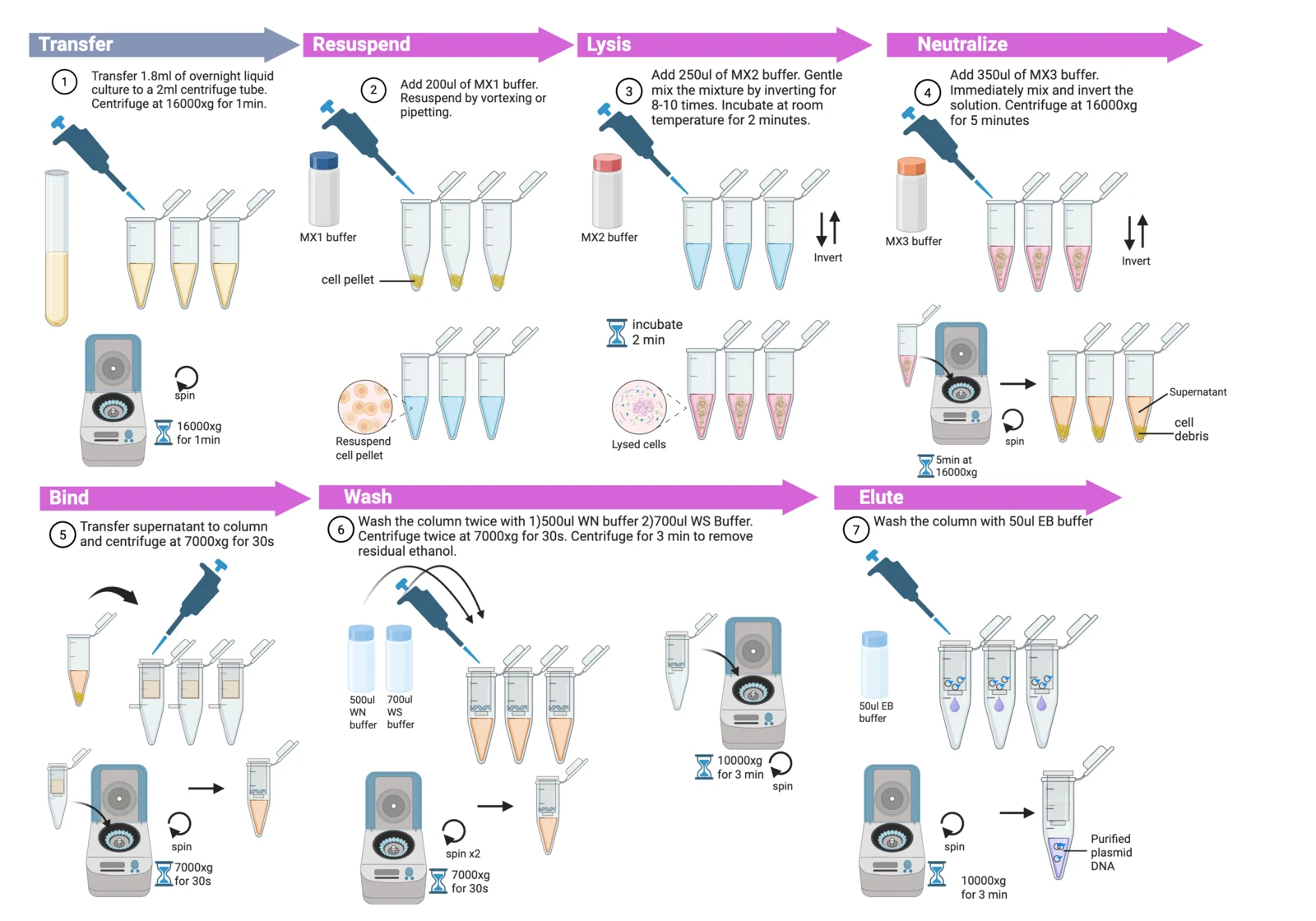
DNA isolationIsolate plasmid DNA from bacterial cultures for concentration measurement and downstream applications.
Recombinant protein production
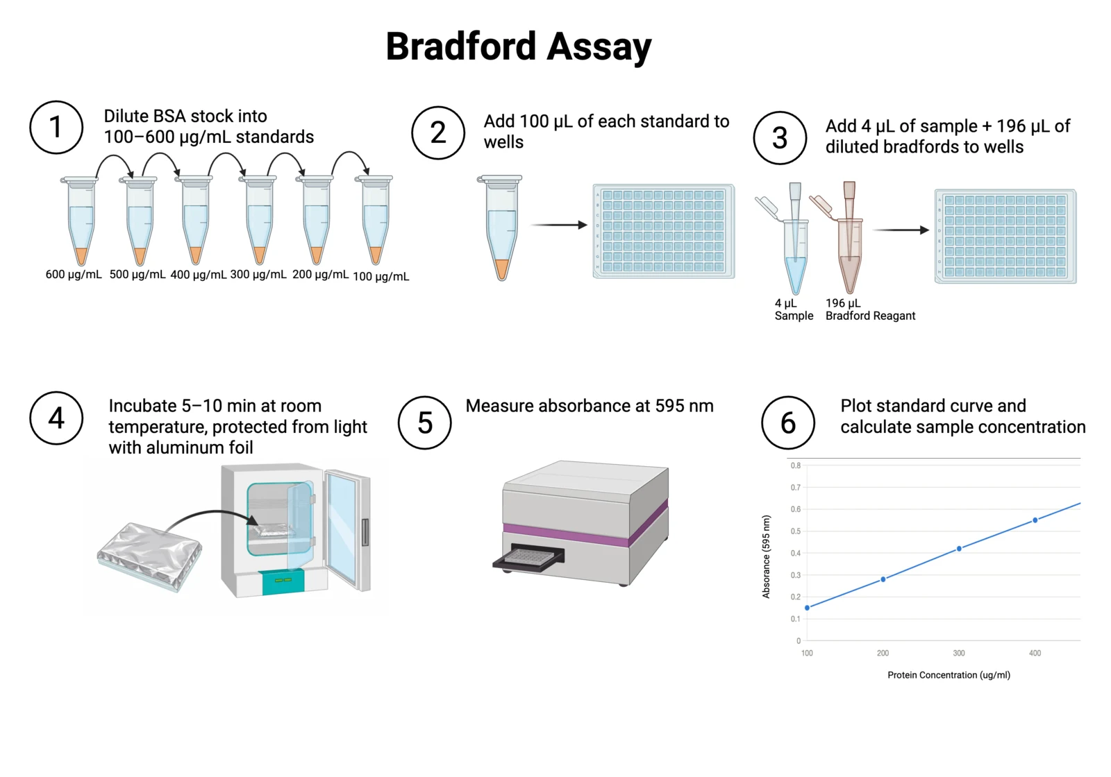
Bradford protein assayA colorimetric method for quantifying protein concentration in a sample.
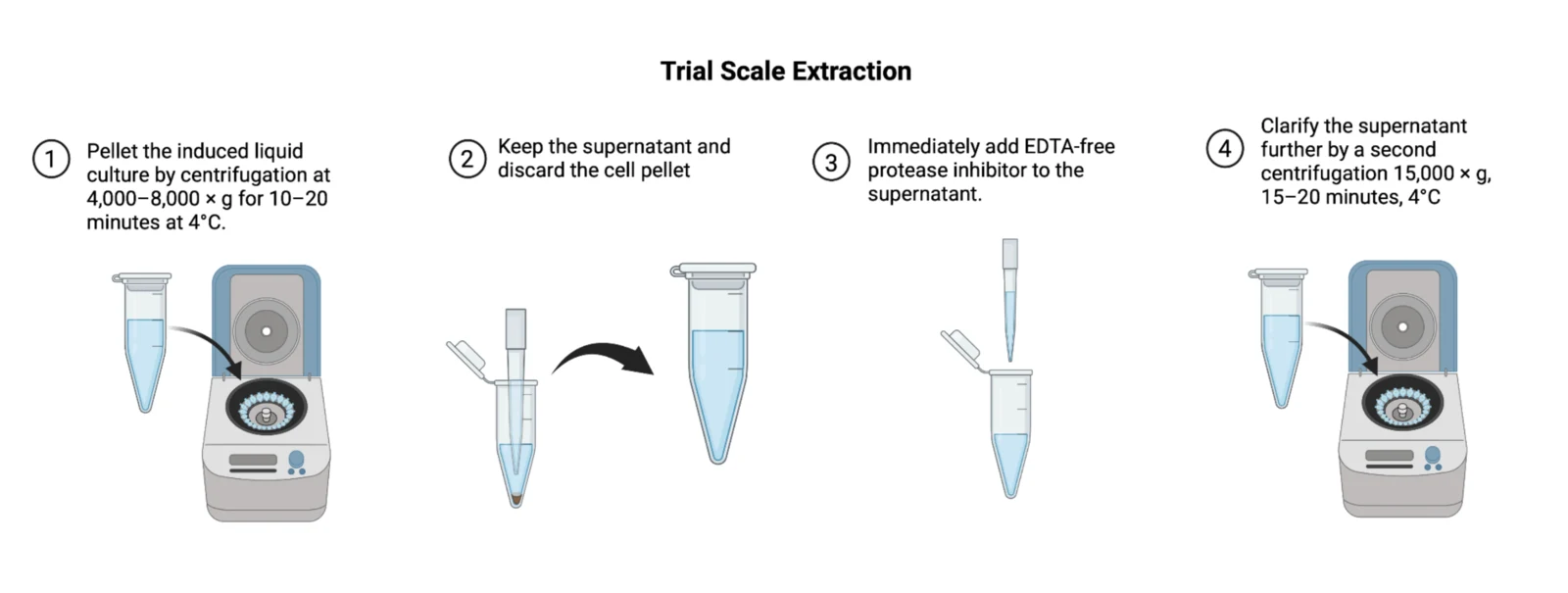
Trial scale protein extractionEvaluate protein extraction conditions on a small scale before proceeding to large-scale production.
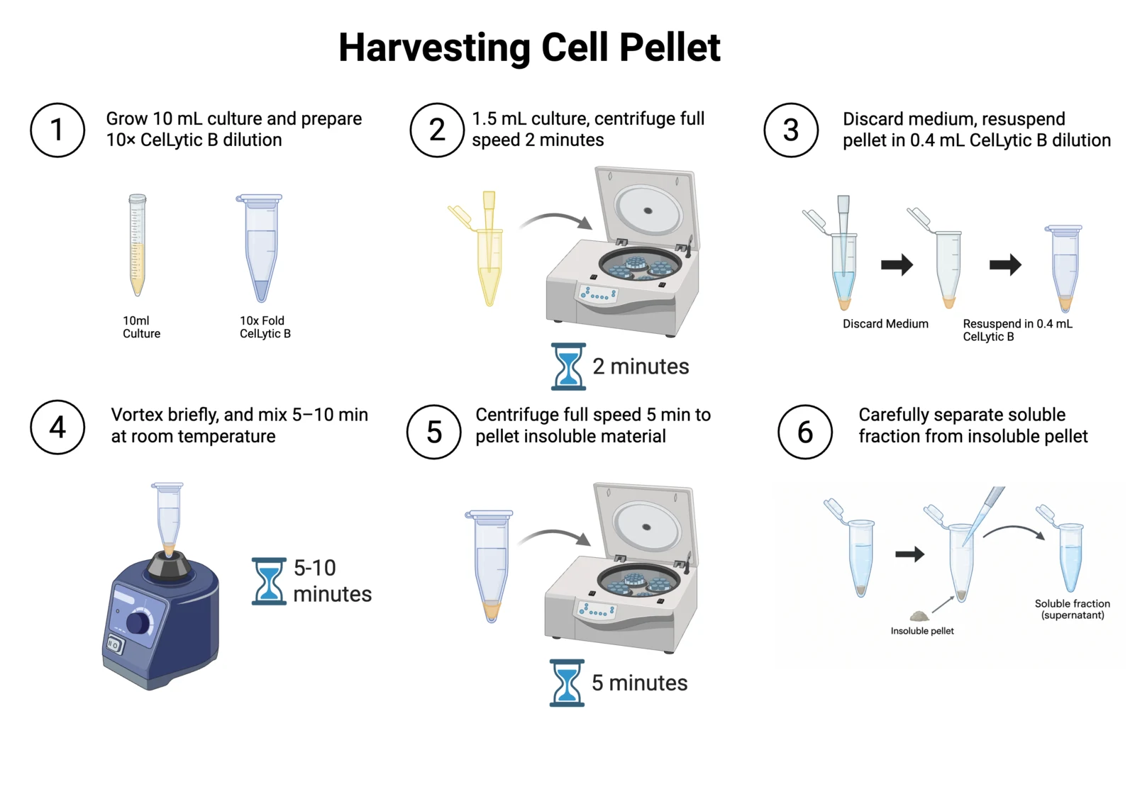
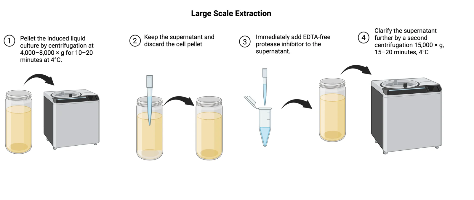
Large scale protein extractionAfter establishing suitable conditions on a small scale, scale up the bacterial culture to produce a larger amount of the target protein.
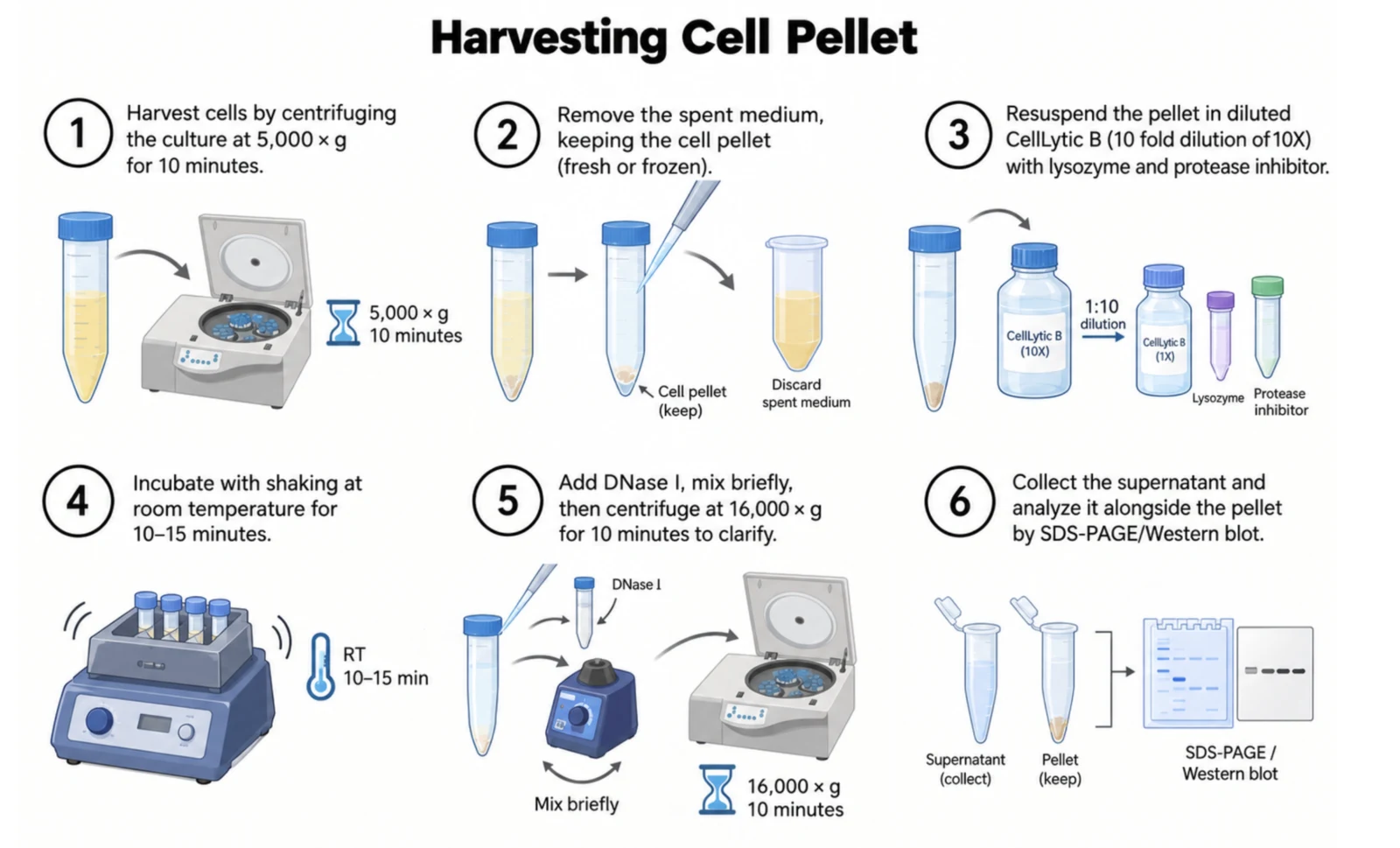
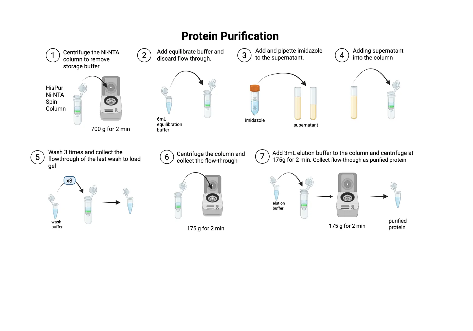
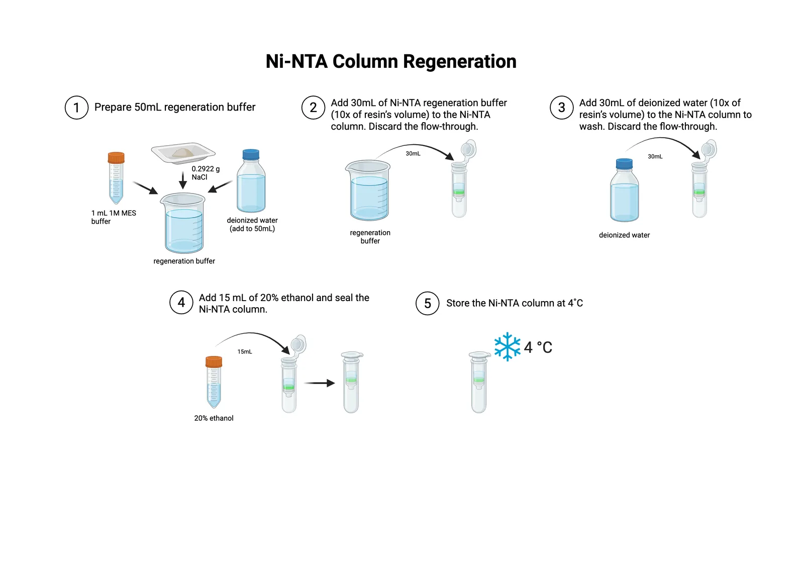
Protein purification and dialysisTo remove residual salts and concentrate the desired protein following large-scale production.
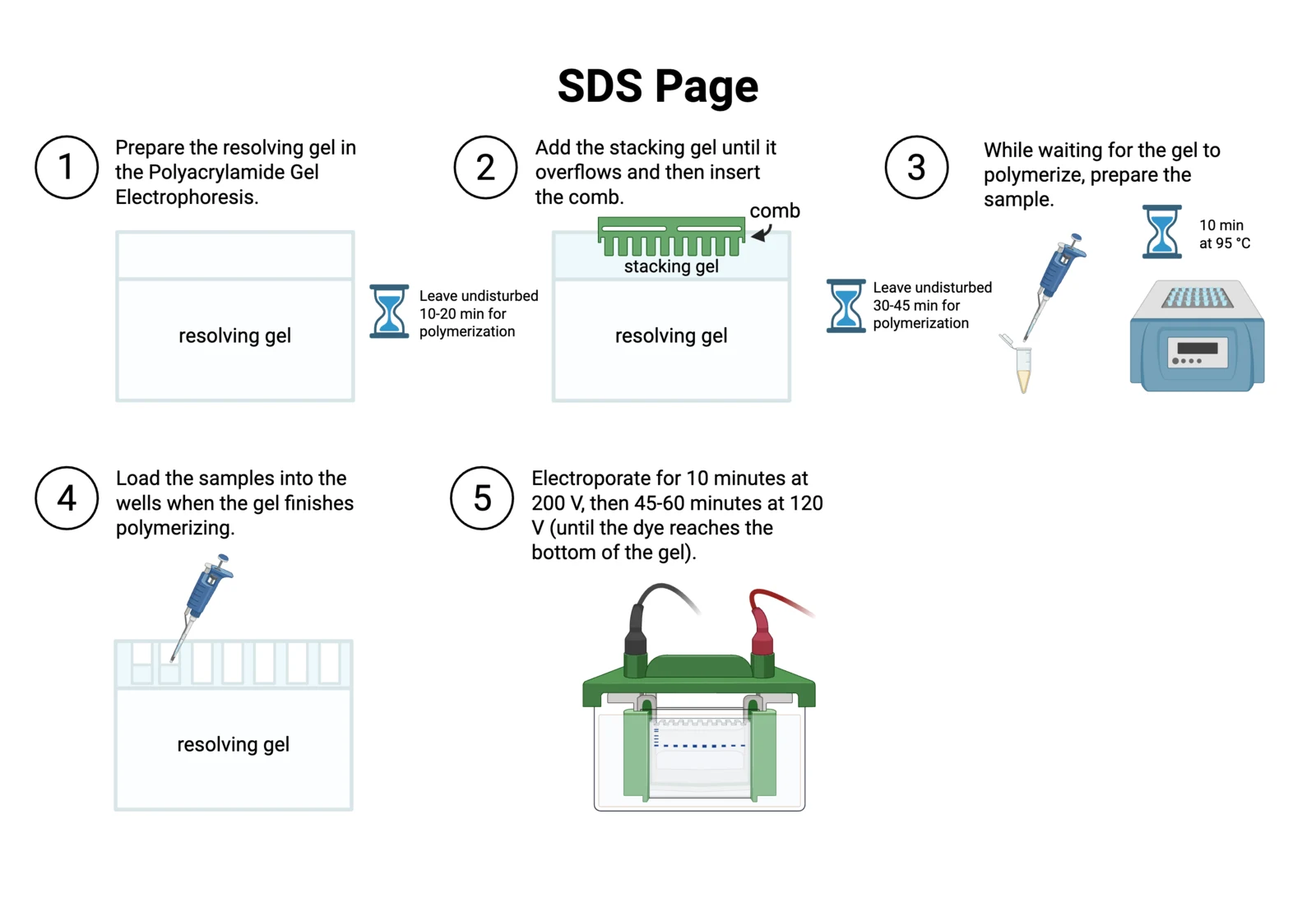
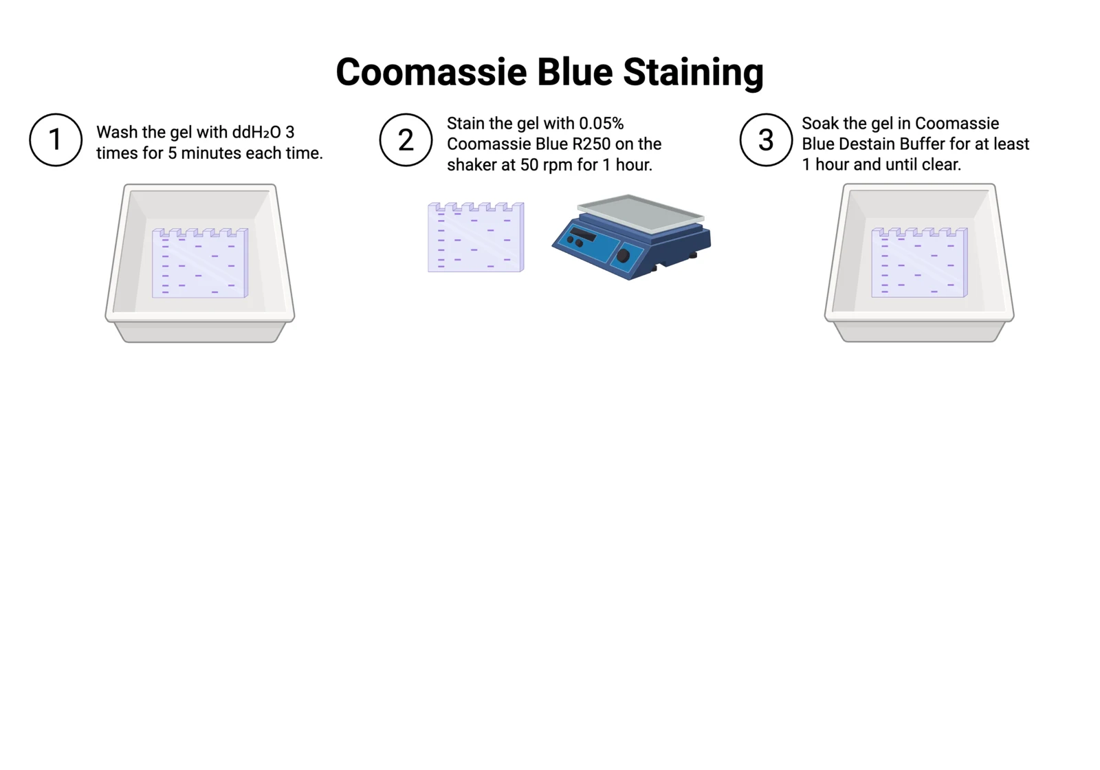
SDS-PAGE and Coomassie stainingTo separate proteins based on their molecular weight using polyacrylamide gel electrophoresis.
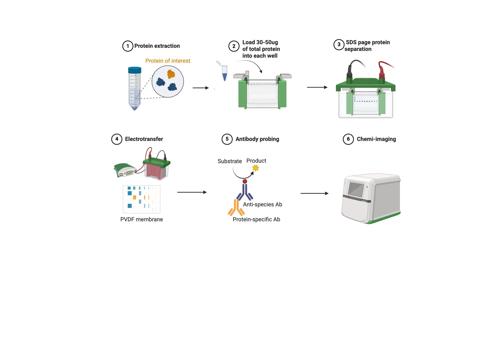
Western blottingDetect a His-tagged protein on a PVDF membrane with an anti-His primary antibody and chemiluminescence.
DNPH assay for ACC deaminase activityEstablish a microscale DNPH assay for determining ACCD activation levels, by measuring α-ketobutyrate accumulation and assessing strain performance across cofactor conditions and gene constructs.
Plant stress assays
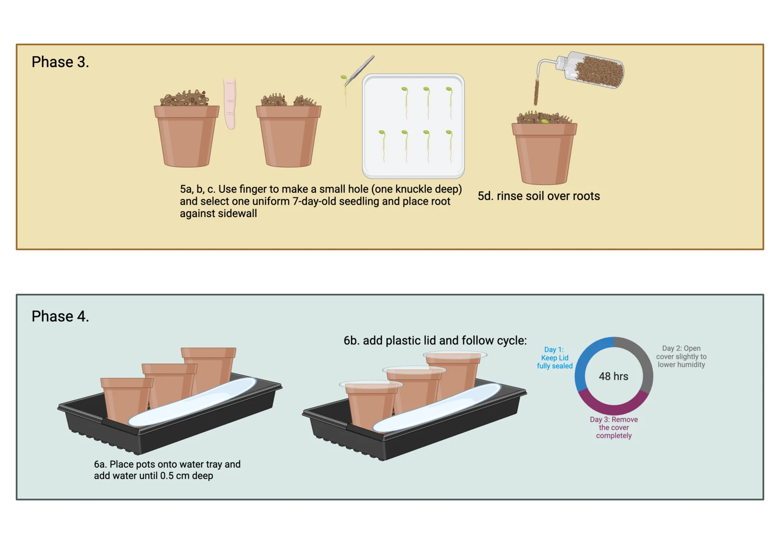
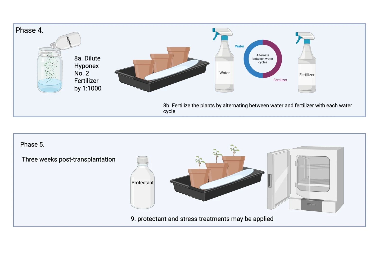
Arabidopsis in soilCultivate Arabidopsis seedlings on soil pots with a standard protocol.
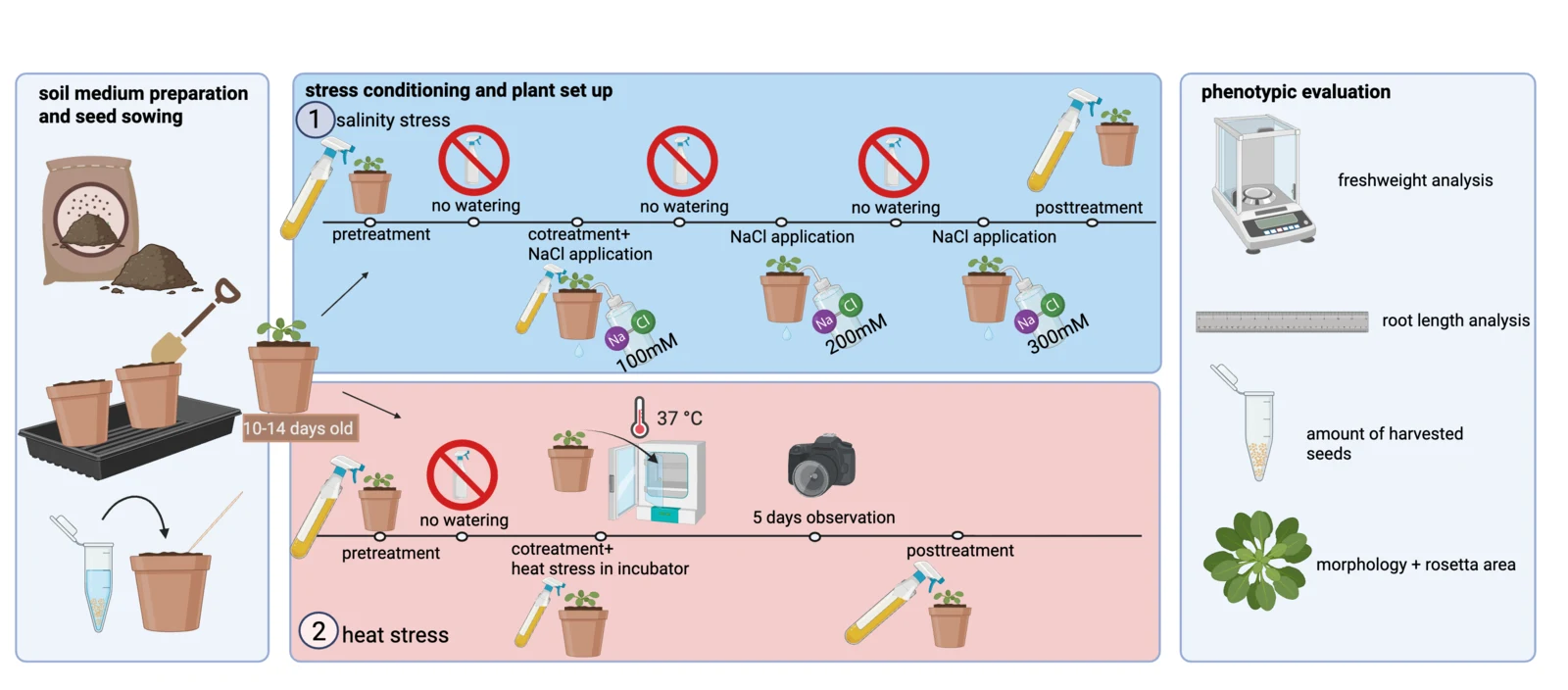
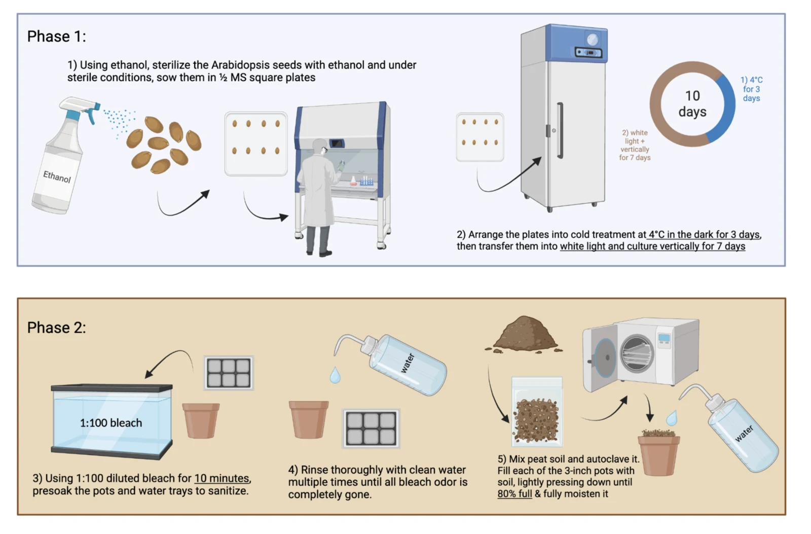
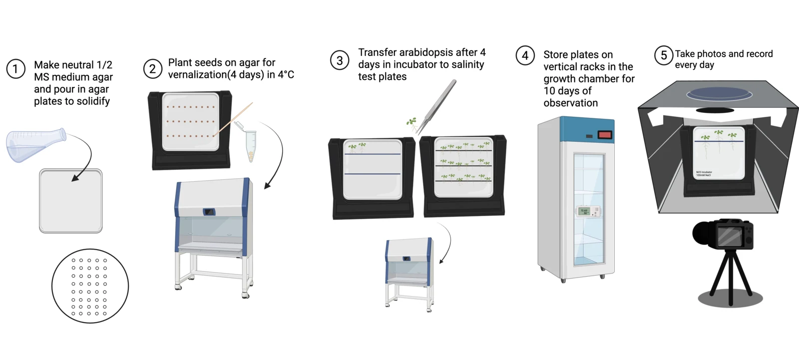
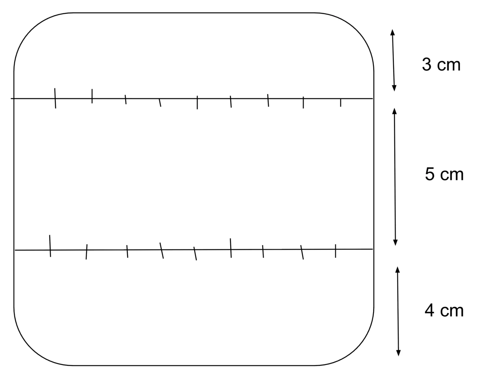
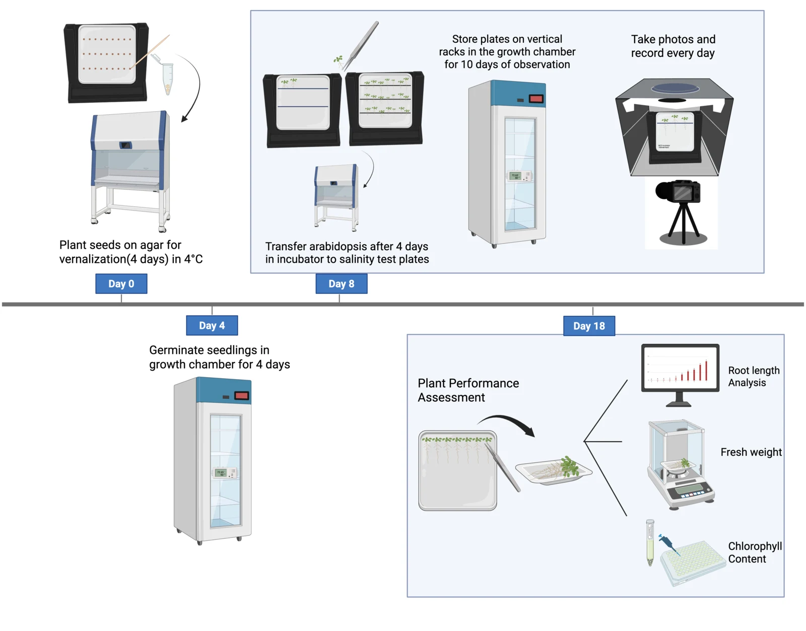
Salinity stress on agarApplying various salinity stress levels to Arabidopsis on agar to evaluate which concentration of trehalose protectant provides the best rescue effectiveness.
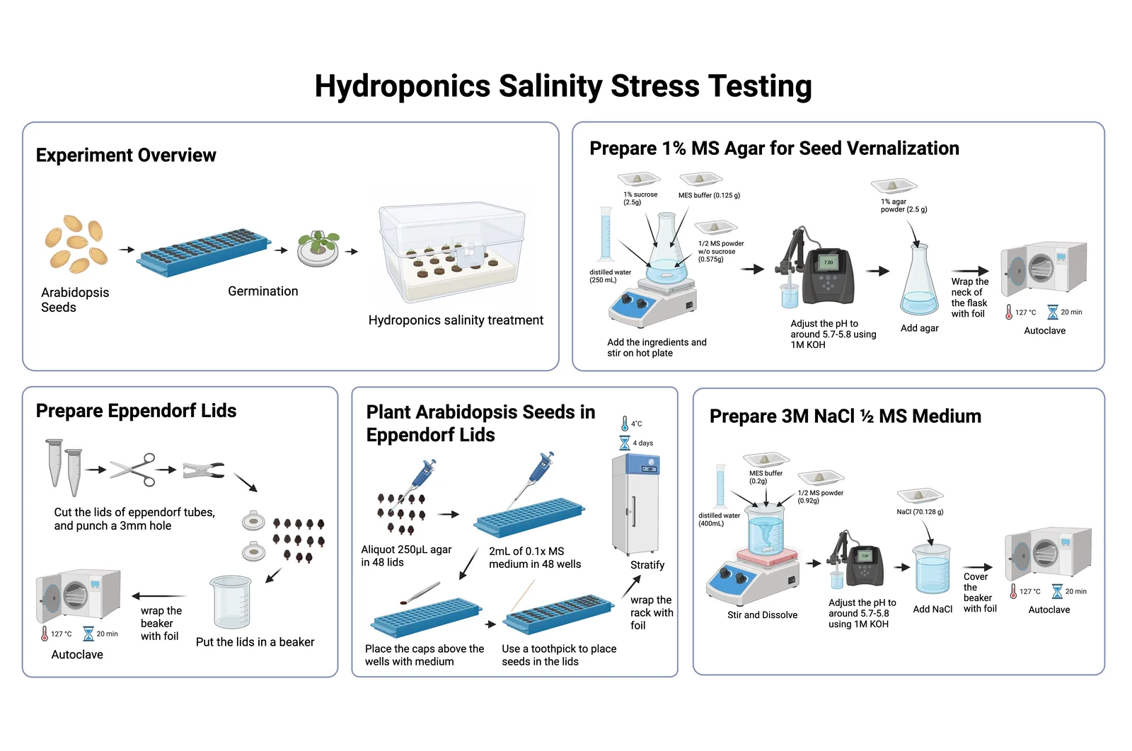
Salinity stress on hydroponicsApplying salinity stress at different time periods to Arabidopsis on hydroponics to evaluate whether our protectant are effective at rescuing Arabidopsis.
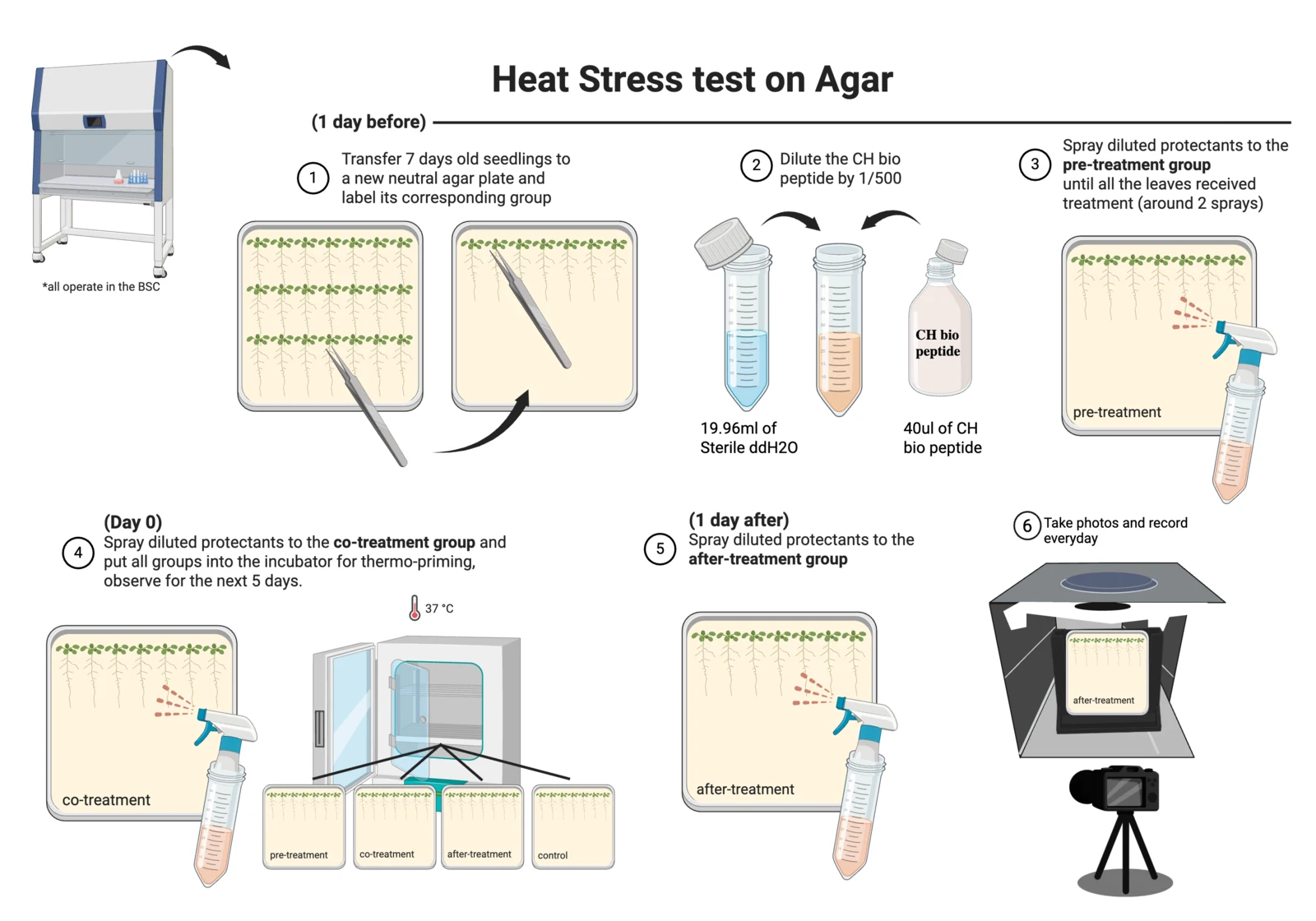
Heat stress on agarApplying heat stress to Arabidopsis on agar to evaluate which treatment time of CH bio peptide provides the best rescue effectiveness.
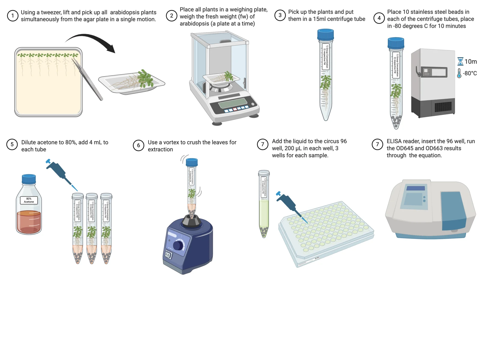
Chlorophyll extractionExtract chlorophyll of each set of Arabidopsis groups after plant experimental trials for readout data and analysis.
Bioreactor and measurement
SiO2 OD600 calibration of the inline photometersBuild a particle concentration vs. OD600 regression curve by cumulative titration of a known-concentration monodisperse silica (SiO2) microsphere suspension.
Bioreactor perfusion runCulture in the hollow fibre module, with the protectant collected on the shell side.
- Hardware → Bioreactor: how the module, pump and photometers are built and run
- Measurement → Bioreactor: the photometer, pressure and flow numbers and their state
- Safety → Bioreactor safety: the shell-side sterility plates that test containment
References
- Arnon DI. Copper enzymes in isolated chloroplasts. Polyphenoloxidase in Beta vulgaris. Plant Physiology 1949;24(1):1-15.
- Bradford MM. A rapid and sensitive method for the quantitation of microgram quantities of protein utilizing the principle of protein-dye binding. Analytical Biochemistry 1976;72:248-254.
- Viogene. Mini Plus Plasmid DNA Extraction System handbook.
- Thermo Fisher Scientific. HisPur Ni-NTA Spin Purification Kit and Slide-A-Lyzer G3 Dialysis Cassettes instructions.
- Sigma-Aldrich. CelLytic B Cell Lysis Reagent product information.
- Bio-Rad. Gene Pulser Xcell Electroporation System instruction manual.
- Penrose DM, Glick BR. Methods for isolating and characterizing ACC deaminase-containing plant growth-promoting rhizobacteria. Physiologia Plantarum 2003;118(1):10-15.
- Beal J, Farny NG, Haddock-Angelli T, et al. Robust estimation of bacterial cell count from optical density. Communications Biology 2020;3:512.
- van de Hulst HC. Light Scattering by Small Particles. New York: John Wiley & Sons; 1957.